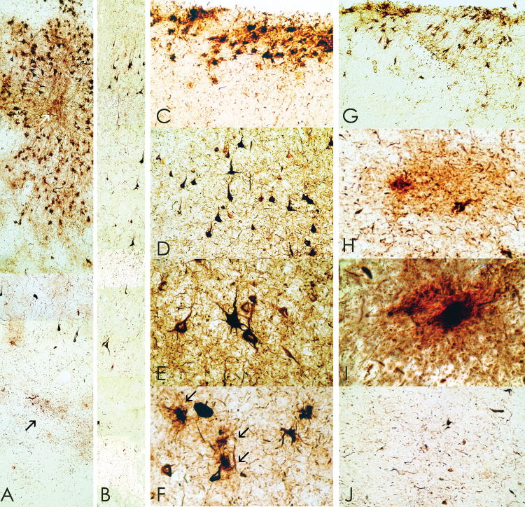
Figure 3.
Whole mount 50-μm sections from cases 1 and 2 immunostained with anti-tau monoclonal antibody AT85. (A) Case 2. There is a prominent perivascular collection of neurofibrillary tangles (NFTs) and astrocytic tangles evident in the superficial cortical layers with lesser involvement of the deep laminae. Prominent neuropil neurites (NNs) are found in the subcortical U-fibers (arrow), original magnification x150. (B) Case 1. There is a preferential distribution of NFTs in layer II and NNs extending into the subcortical white matter even in mildly affected cortex, original magnification x150. (C) Case 1. Focal subpial collections of astrocytic tangles and NFTs are characteristic of chronic traumatic encephalopathy (CTE), original magnification x150. (D) Case 1. The shape of most NFTs and NNs in CTE is similar to those found in Alzheimer disease, original magnification x150. Some NFTs have multiple perisomatic processes (E) and spindle-shaped and dot-like neurites are found in addition to thread-like forms (E, Case 1, original magnification x350. (F) Case 1. Astrocytic tangles are interspersed with NFTs in the cortex (arrows, original magnification x350). (G) Case 1. Tau-immunoreactive astrocytes are common in periventricular regions, original magnification x150. (H, I) Case 1. Tau-immunoreactive astrocytes take various forms; some appear to be protoplasmic astrocytes with short rounded processes (H, I, double immunostained section with AT8 (brown) and anti-glial fibrillary acidic protein (red), (H) original magnification x350, (I) original magnification x945). (J) Case 2: Dot-like or spindle-shaped neurites predominate in the white matter although there are also some threadlike forms, original magnification x150.

