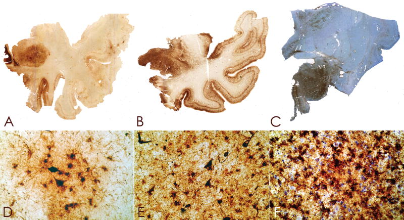
Figure 4.
(A–C) Whole mount 50-μm-thick coronal sections immunostained for tau (AT8) from case 1 (A), case 2 (B), case 3 (C) (counterstained with cresyl violet) showing extremely dense deposition of tau protein in the amygdala with increasing severity from left to right. (D–F) Microscopically, there is a moderate density of NFTs and astrocytic tangles in case 1 (D), the density is increased in case 2 (E), and extremely marked in case 3 (F), original magnification x350.

