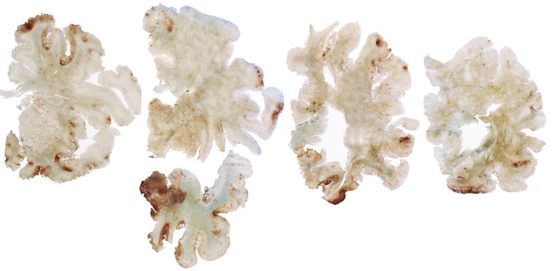
Figure 5.
Whole mount 50-μm coronal sections of case 2 (A) and case 3 (B), immunostained for tau (AT8) and counter-stained with cresyl violet. There is extremely dense deposition of tau protein in the hippocampus and medial temporal lobe structures. There is also prominent tau deposition in the medial thalamus.

