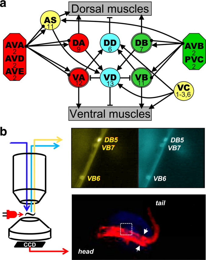
Figure 1.

Locomotor neuronal circuit and recording set up. a, Schematic of connectivity of the locomotor circuit [based on Chalfie and White (1988) and Von Stetina et al. (2006) with additional connections extracted from Altun and Hall (2009) and supplemental material of Chen et al. (2006)]: five pairs of interneurons (octagons) innervate eight groups of motoneurons (circles) that innervate either dorsal or ventral muscle of the body wall. Connecting lines represent excitatory (arrows) or inhibitory (T lines) synapses and gap junctions (solid circles). The number of neurons in each group is indicated. DB, VB, and VA, which were recorded from in this study, have a double-line frame. For simplicity, connectivity among interneurons is not presented and interneurons are grouped based on targets. b, Recording set-up. Epifluorescence calcium imaging of motoneurons was done through a water-immersion objective on an upright microscope. An example of pseudocolored yellow and cyan channels is shown in the upper right panel. The nematode's behavior was imaged through the condenser using a modified webcam. The animal was illuminated with a red LED positioned close to the recording chamber. An image of the nematode viewed this way is shown in the lower right hand panel. The arrows indicate the caudal limit of the glued region and the field of view of the calcium imaging is shown by the white rectangle.
