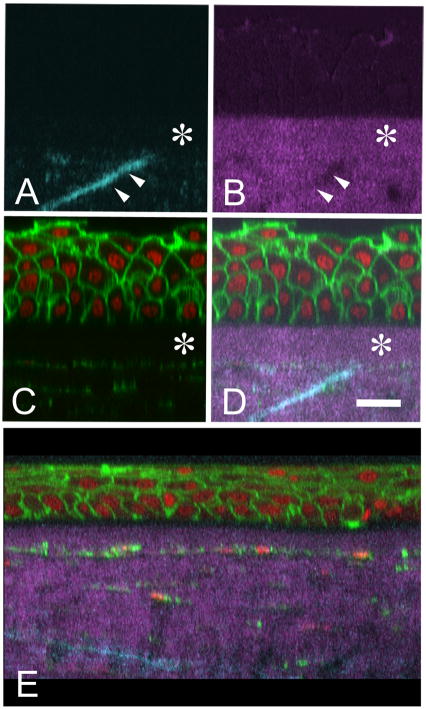
Figure 1.
Reconstructed cross-sectional images of normal human cornea showing forward (A. cyan), backward (B, magenta) SHG signal co-localized with Phalloidin to detect actin and Syto-59 to detect nuclei (C, Green and Red respectively). Co-localized image (D) shows that SHG imaging can detect the anterior limiting lamina (Bowman’s layer, asterisk) and the insertion of ‘sutural’ collagen fibers into the anterior limiting lamina (double arrows). Cross-sectional reconstruction of a keratoconus cornea (E) shows the absence of ‘sutural’ collagen fibers. Bar = 20 μm (Taken from Figure 4, J Cataract Ref Surg 32:1784, 2006.)

