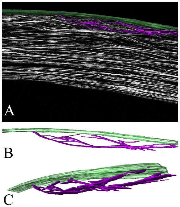
Figure 5.
3-D reconstructed sutural lamellae in the human cornea. (A) 1 mm central corneal region from a HRMac corneal cross-section after segmentation in Amira. Bowman’s layer is rendered in green, the surtural lamellae are purple. (B) and (C) show the extracted sutural lamellae rendering in two different rotations. These images can be panned, zoomed and rotated at will. Taken from Winkler et al, High resolution macroscopy (HRMac) of the eye using non-linear optical imaging. Proceeding of SPIE, Vol 7589: 758906.

