Figure 7.
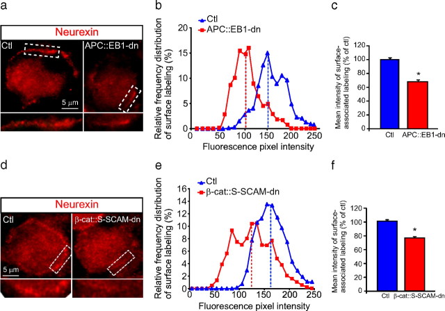
a–f, Neurexin surface clusters are decreased on presynaptic terminals that contact postsynaptic neurons expressing APC::EB1-dn or β-cat::S-SCAM-dn. a, d, Micrographs of immunofluorescence double-labeled E13 CG frozen sections showing that Nrx surface clusters (red; a, d) are decreased on presynaptic terminals that contact APC::EB1-dn (a) and β-cat::S-SCAM-dn (d) neurons compared with Ctl neurons. Insets, twofold magnification views of boxed regions. b, c, e, f, Nrx staining shows shifts to lower pixel intensity levels (b, e) as well as 31.5% and 22.6% reductions in mean intensity levels (c, f) at synapses on APC::EB1-dn (b, c)- and β-cat::S-SCAM-dn (e, f)-expressing neurons, respectively, relative to Ctl neurons. (APC::EB1-dn: *p < 6.9 × 10⁻⁹, Student's t test, n = 21 DN and 14 Ctl neurons; β-cat::S-SCAM-dn: *p < 5.2 × 10⁻²² Student's t test, n = 22 DN and 21 Ctl neurons). Dashed vertical lines indicate the median intensity values (b, e). Bars represent the mean ± SEM (c, f).

