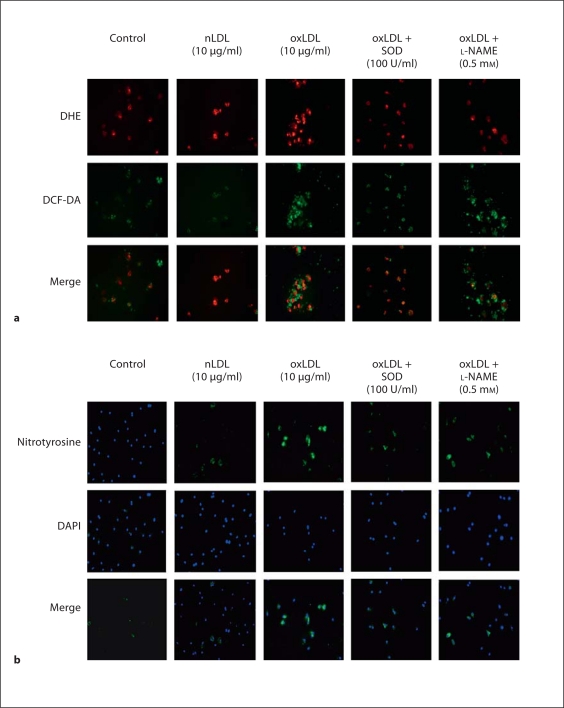
Fig. 3.
Effects of nLDL and oxLDL on reactive oxygen species in wild-type EPC. EPC cultures were exposed to nLDL (10 μg/ml), oxLDL (10 μg/ml), oxLDL (10 μg/ml) + SOD (100 U/ml) or oxLDL (10 μg/ml) + L-NAME (0.5 mM) in serum-free medium. The control well was treated with medium alone. a Quantification of DCF and DHE staining in EPC. Data are presented as mean ± SD, n = 3; + p < 0.05 oxLDL versus nLDL or control; * p < 0.05 versus oxLDL. b Nitrotyrosine staining for protein nitrosylation. Nitrotyrosine is stained green. The slide was counterstained with DAPI to detect nuclei, which appears blue. This set of immunofluorescent photomicrographs is from a single experiment and are representative of three experiments.

