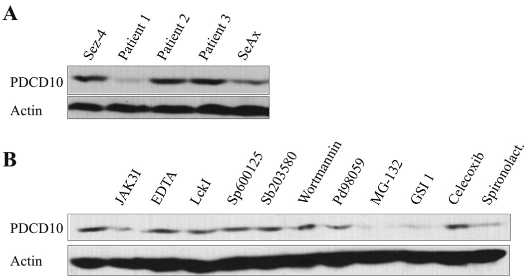
Figure 1. Malignant SS cell lines and in primary leukemic Sézary cells constitutively express PDCD10.
(A)Western blot analysis of whole-cell lysates from SS cell lines established from peripheral blood of patients diagnosed with SS: Sez-4 (lane 1) and SeAx (lane 5), and from primary cells obtained from peripheral blood of three patients (patient 1–3) diagnosed with SS (Lane 2–4). The blots were probed with the antibodies against PDCD10 and visualized as bands at ~26kDa. (B) Malignant SS cells (SeAx) were treated with JAK3I (3 µM), EDTA (5 µM), LckI (10 µM), Sp600125 (5 µM), Sb203580 (10 µM), Wortmannin (5 µM), PD98059 (50 µM), MG-132 (10 µM), GSI 1 (5 µM), Celecoxib (50 µM), spironolactone (3 µM) or the vehicle (DMSO) for sixteen hours prior to Western blot analysis for the expression of PDCD10.

