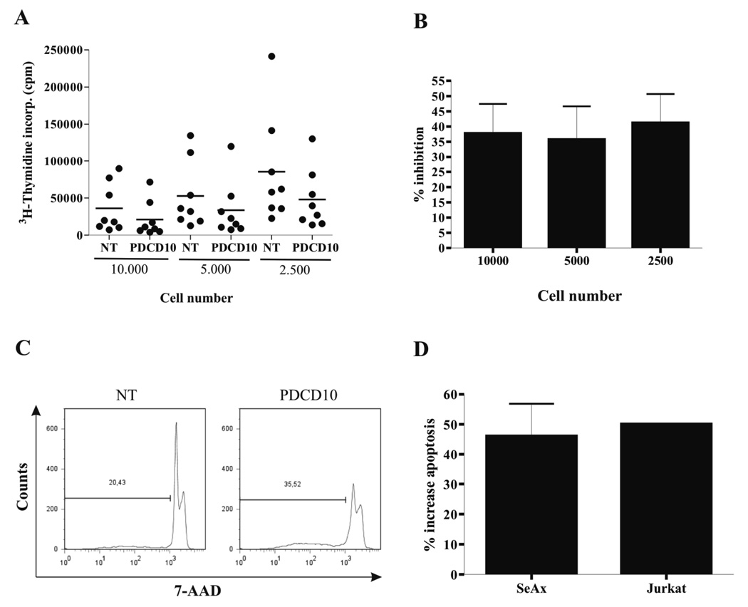
Figure 5. Specific downregulation of PDCD10 inhibits proliferation and induces apoptosis.
(A) Dot blot and (B) % inhibition of 3H-thymidine incorporation of malignant SS cells (SeAx) that were transiently transfected with non-targeting (NT) or PDCD-10-specific siRNA. 10.000, 5.000 or 2.500 transfected cells/well were cultured for 48 hours in a 96 round bottom culture plate. Sixteen hours before harvest, 3H-thymidine (1 µCi ~[0.037 MBq]/well) was added to the cultures and the results are expressed as mean counts per minute of eight independent experiments. (38% inhibition, p < 0.001 at 10.000 cells per well, 36 % inhibition, p < 0.05 at 5.000 cells per well, 48 % inhibition, p < 0.001, at 2.500 cell per well, Wilcoxon Rank sum test for paired differences). (C) SeAx cells were stained with 7-AAD and analyzed by flow cytometry. Data are representative of five independent experiments. (D) % increase in apoptosis of malignant SS cells (SeAx) and Jurkat cells 48 hours after transient transfection with non-targeting (NT) or PDCD10-specific siRNA. Error bars of SeAx represent SEM of five independent experiments while measurement of apoptosis in the Jurkat cell line following transfection only was performed once.

