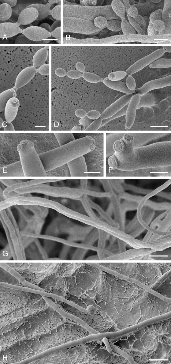
Fig. 56.
Cladosporium perangustum (CBS 125996). A. Conidia with very gentle surface ornamentation showing irregularly reticulate structures. B. A coherent view on conidiophores, stipes, aerial hyphae and conidia. C. Secondary ramoconidia, conidia and scars. The conidia at the upper right show some cell wall structures. D. Conidiophore with secondary ramoconidia, intercalary and small terminal conidia. Note the disruptions of the cell walls between the conidia. E. Scars on very elongated secondary ramoconidia. F. Scar-pattern at the end of the conidiophores. Note the flattened separation domes. G. Ropes of aerial hyphae. H. Running segmented hyphae that may form conidiophores and not segmented aerial hyphae. Note the blastoconidium on one hypha. Scale bars = 2 (A, C, E–F), 5 (D, G), 10 (B, H) μm.

