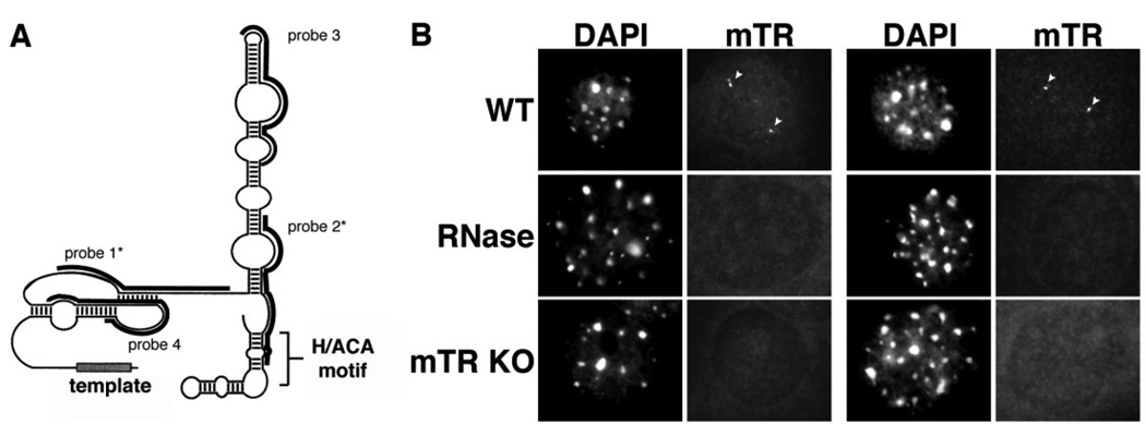
Figure 1. Mouse telomerase RNA is found in small spherical foci within the nuclei of cultured mouse cell lines.
A. Schematic structure of mTR. The predicted secondary structure of mTR is shown [70]. Black bars indicate the regions encompassed by each oligonucleotide probe. Asterisks denote the two probes (probes 1 and 2) used throughout this manuscript. B. FISH procedure specifically detects the presence of mTR. mTR FISH was performed on wild type MEF cells (WT, RNAse panels) or MEF cells derived from mTR −/− mice (mTR KO panels). Arrowheads denote intranuclear mTR foci present in the WT cells (WT panels), which are lost upon treatment of cells with RNAse A prior to FISH (RNAse panels). DAPI was used as a nuclear stain. Scale bar, 10 microns.

