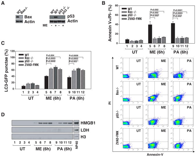
Figure 3. HMGB1 release and autophagy is detected in the absence of measurable apoptosis.
(A) Immunoblots are shown for Bax and p53 knockout in HCT116 cells. (B-D) WT, Bax knock out, p53 knockout or pan-caspase inhibitor treated (ZVAD-FMK, 20 μm) HCT116 cells were treated with melphalan, “ME”, 160 μg/ml or paclitaxel, “PA”, 10 μg/ml for 6 h. and then assayed for measures of early apoptosis (annexin V+/PI−) by flow cytometry (B), autophagy by quantification of the percentage of cells with GFP-LC3 punctae (C) and HMGB1 release by western blot analysis (LDH and H3 were both used as controls for protein leakage from damaged cells) (D). Representative western blots of the indicated proteins are presented.

