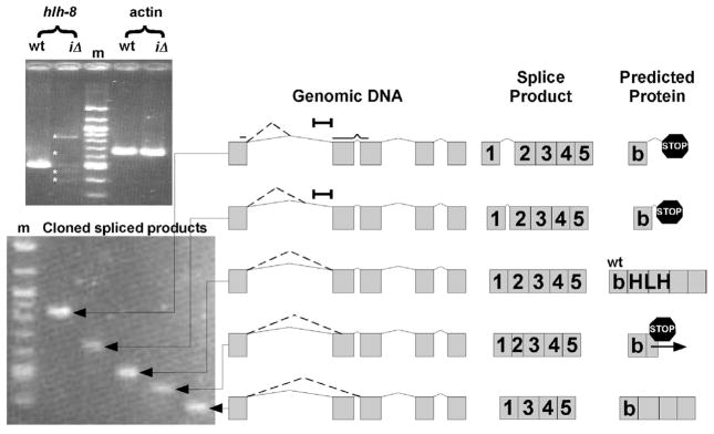
Figure 8. Splicing defects in hlh-8(iΔ) animals.
RT-PCR revealed 4 alternate spliced products (asterisks) of hlh-8 in hlh-8(iΔ) animals (Top Gel). Individual clones of spliced fragments (Bottom Gel). The schematics on the right are drawn to scale. ‘Genomic DNA’ indicates where splicing occurs in each fragment (dotted line). Exons are represented by gray boxes and introns by solid lines. A black bar indicates the region of DNA removed in hlh-8(iΔ) animals. ‘Spliced Product’ designates the various splice products determined from sequencing data. ‘Predicted Protein’ depicts the polypeptide that results from each of the spliced products. The third splice product is the wild-type product, identified by ‘wt’. Premature stop codons (stop signs) and the frameshift (horizontal arrow) are indicated.

