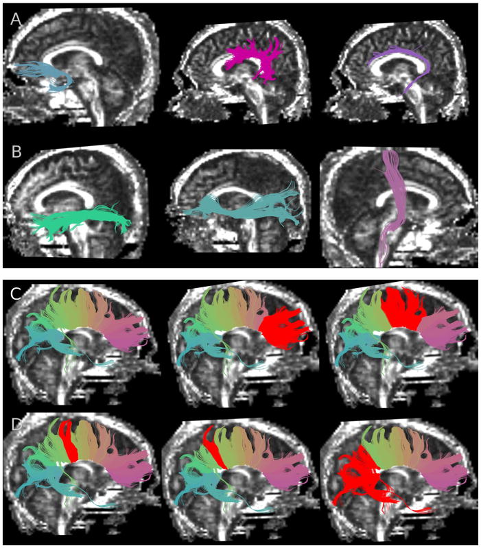
Fig. 1.
Segmented white matter fiber tracts superimposed on fractional anisotropy images. (A) From left to right: uncinate fasciculus, arcuate fasciculus, cingulum bundle; and (B) from left to right: inferior longitudinal fasciculus, inferior occipitofrontal fasciculus, corticospinal tract; (C) from left to right: corpus callosum (whole structure with fibers color-coded according to similarity of shape and location), genu of corpus callosum (CC1) selected in red, premotor and supplementary motor fibers of corpus callosum (CC2) selected in red; and (D) from left to right: motor fibers of corpus callosum (CC3) selected in red, sensory fibers of corpus callosum (CC4) selected in red, and parietal, temporal and occipital fibers of corpus callosum (CC5) selected in red.

