
Figure 1.
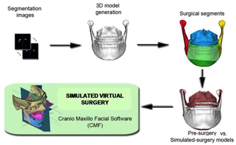
Sequence of image analysis procedures used for virtual surgical simulation: After segmentation of anatomic structures, i.e. outlining the shape of structures visible in the cross-sections of a CBCT volumetric dataset, the virtual cuts were performed. For each patient, simulated surgery outcomes were created, to compare to presurgery and actual surgery models. Virtual cuts matched clinical osteotomy segments that in this example were: chin, left ramus, right ramus, mandibular body and/or maxillary body. The virtual surgical segments were then displaced to determine if virtual surgery performed on the Cone beam CT surface models can correctly simulate the actual surgical outcome.