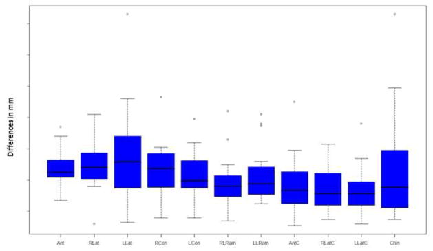
Figure 3.
The differences between virtual and actual post-surgery models are shown below. The x axis shows the 11 regions of interest and the y axis shows the difference in mm between the two images. All regions of interest except the left lateral maxilla showed a mean and median difference less than the 0.5 mm spatial resolution of the acquired image. (Ant = Anterior Maxilla, RLat = Right lateral maxilla, LLat = Left lateral maxilla, RCon = Right condyle, LCon = Left condyle, RLRam = Right lateral ramus, LLRam = Left lateral ramus, AntC = Anterior Corpus of the mandible, RLatC = Right lateral corpus of the mandible, LLatC = Left lateral corpus of the mandible, Chin = Chin)

