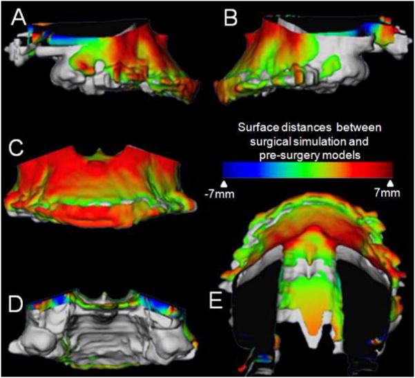
Figure 6.
Example of a maxillary impaction case in which surgical simulation helped plan areas and amount of bone removal for impaction. Superimposition of maxillary segment of virtual surgery models and pre-surgery models of patients treated with maxillary advancement and impaction. A, Right lateral view, B, Left lateral view, C, Frontal view, D, Posterior view and E, Superior view. The grey image is the pre-surgery model and the image with the color map is the post virtual (simulated) surgery image. Color maps demonstrate the location, direction, and magnitude of the differences between these models. Note the dark blue area in the posterior part of the maxilla indicating that 7mm of posterior bone removal will be necessary during the surgery.

