
Figure 1.
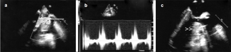
Intraoperative transesophageal echocardiogram following AVR showing turbulent abnormal flow in the LVOT (a, arrow heads) with a dynamic peak gradient of 58 mmHg (b) and a small left ventricle with thick basal sigmoid shaped septum (c, arrow heads) causing narrow LVOT; LV, left ventricle