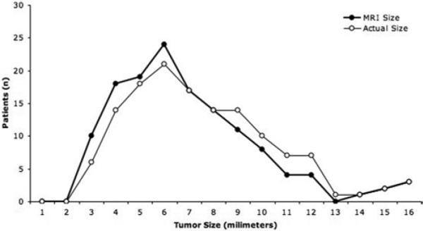
Fig. 3.

Graphic representation of tumor size on MR imaging (bold black line) compared with the tumor size at resection. Magnetic resonance imaging–based measurement was within 2 mm of the tumor size at surgery in most cases, although there was an overall tendency to underestimate tumor size on MR imaging, a factor that is probably related to the resection of the pseudocapsule with the adenoma.
