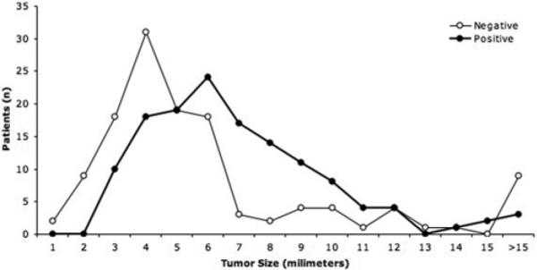
Fig. 4.

Graphic representation of tumor size in patients with negative MR imaging compared with those in patients with positive MR imaging (closed circles). The patients with negative MR imaging (open circles) tended to have smaller tumors, although there is considerable overlap in the range of tumor size with and without detection on MR imaging.
