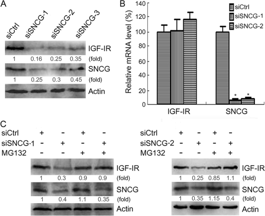
FIGURE 6.
SNCG prevents proteasomal degradation of IGF-IR. A, HepG2 cells were transfected with siCtrl or three siRNA duplex to SNCG that target different sites within SNCG mRNA (siSNCG, siSNCG-2, siSNCG-3). Forty-eight hours later the total proteins were harvested and subjected to Western blot analysis of IGF-IRβ and SNCG expression. Forty-microgram aliquots of total proteins were loaded. The relative content of IGF-IR and SNCG after normalization to actin is shown. B, HepG2 cells were transfected with siCtrl, siSNCG, or siSNCG-2. Twenty-four hours later the total RNA were isolated and subjected to quantitative real-time RT-PCR analysis of SNCG and IGF-IR transcription. β-Actin served as a reference gene. Four replicates were tested in each group. *, p < 0.001, compared with siCtrl. There was no significant difference in the levels of IGF-IR transcripts. C, HepG2 cells were transfected with siCtrl, siSNCG-1, or siSNCG-2 and treated with or without 5 μm MG132. Forty-eight hours later the total proteins were harvested and subjected to Western blot analysis of IGF-IRβ and SNCG expression. Forty-microgram aliquots of total proteins were loaded. The relative content of IGF-IR and SNCG after normalization to actin is shown. A representative of three independent experiments is shown.

