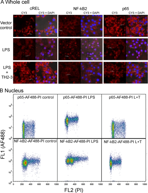
FIGURE 10.
TH2-3 inhibited the LPS-mediated nuclear translocation of NF-κB subunits. RAW264.7 cells were stimulated with 100 ng/ml LPS, LPS co-treated with 100 μg/ml TH2-3, or an untreated vector control for 1 h. A, cells cultured over the coverslip were fixed in 4% paraformaldehyde, and localizations of p65, NF-κB2 and cREL were observed by probing with a respective polyclonal anti-rabbit antibody and Cy3-conjugated secondary antibody. Localization of NF-κB alone (Cy3) and co-localization of NK-κB with nuclei stained with DAPI (Cy3 + DAPI) are shown for comparison. Results are representative of three independent experiments. B, flow cytometric analysis of p65 and NF-κB2 expressions in nuclei is shown. Nuclei were isolated from 1-h-treated cells and stained with p65-AF488- or NF-κB2-AF488-conjugated primary antibodies and 10 μg/ml propidium iodide (PI). Then cells were analyzed in BD FACS caliber, and the shift in florescence intensity is displayed in the figure. The results shown are representative of three independent experiments.

