FIGURE 6.
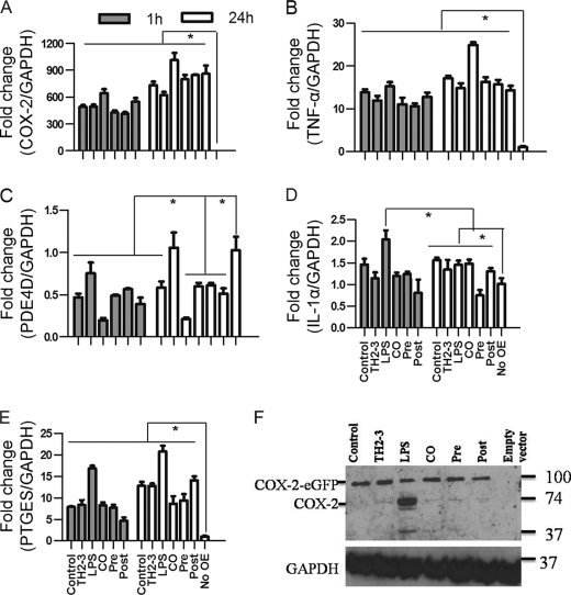
Overexpression of COX-2 induces proinflammatory cytokine expressions in TH2-3-treated cells. RAW264.7 cells were transfected with a pRECEIVER-COX-2-eGFP vector and treated with LPS and/or TH2-3. A–E, the gene expressions of COX-2 (A), TNF-α (B), PDE4D (C), IL-1α (D), and PTGES (E) were measured by a real-time PCR. Multiples of change for each gene were normalized to GAPDH and are relative to the gene expression in unstimulated cells (set to 1) using the comparative Ct method. Values are the mean of three independent experiments (n = 3). Error bars, S.E. *, values differ at p < 0.05. F, Western blot with a COX-2-specific antibody in COX-2-overexpressing cells at 24 h after treatment is shown. Whole cell lysates from three independent experiments were pooled together, and 50 μg of the cell lysate was used to detect the expression.

