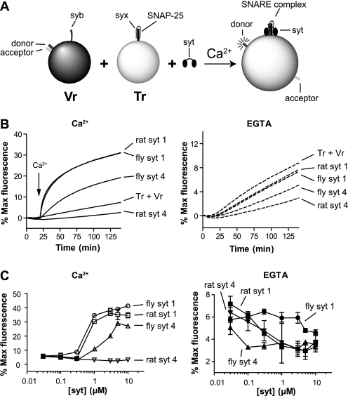
FIGURE 1.
Effect of rat and fly SYT4 on reconstituted SNARE-mediated membrane fusion reactions. A, schematic diagram of the in vitro fusion assay. Tr, t-SNARE vesicle; Vr, v-SNARE vesicle. B, left, the cytoplasmic domain of rat or fly SYT4 (1 μm) was added to SNARE-bearing liposome fusion reactions, and samples were incubated for 20 min prior to the addition of Ca2+. After injection of Ca2+ (1 mm [final], indicated by arrows), fusion was monitored for another 120 min at 37°C. As controls, rat and fly SYT1 were assayed under identical conditions. NBD dequenching signals were normalized to the maximum fluorescence signal, obtained by adding detergent, and plotted as a function of time. Right, experiments were also carried out in the continued presence of 0.2 mm EGTA. C, the final extent of fusion, regulated by SYT1 or SYT4, was plotted against protein concentration in the presence of 1 mm Ca2+ (left panel) or 0.2 mm EGTA (right panel) (n = 3).

