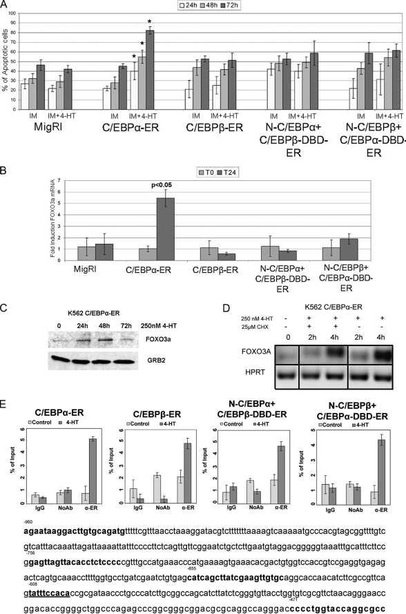
FIGURE 6.
Activation of C/EBPα enhances IM-induced apoptosis of K562 cells. A, shown are the number of apoptotic cells (mean plus S.D., three experiments) in MigRI-transduced and C/EBP-ER-expressing K562 cells after treatment with 0.2 μm IM alone or with 4-HT; * p < 0.05 relative to corresponding 4-HT-untreated sample. B, the histogram shows FOXO3a mRNA levels, assessed by real time Q-PCR, in 4-HT-treated C/EBP-ER-transduced K562 cells. HPRT expression was used as internal control. Error bars denote S.D. of the normalized means of one representative (of two) experiment performed in triplicate. C, shown is expression of FOXO3a in 4-HT-treated C/EBPα-ER K562 cells; expression of FOXO3a was detected by anti-FKHRL/FOXO3a rabbit polyclonal antibody (07-702, UBI). D, FOXO3A mRNA levels, assessed by semiquantitative RT-PCR, in C/EBPα-ER-expressing K562 cells after treatment with 4-HT alone or in the presence of cycloheximide (CHX) are shown. HPRT expression was used as internal loading control. Results are representative of two independent experiments. E, quantitative ChIP assays show binding of C/EBPα, C/EBPβ, and C/EBPα-C/EBPβ chimeric proteins to a segment (nucleotides −655 to −453) of the FOXO3a promoter containing a putative C/EBP binding site (nucleotides −608 to −599) detected by real time Q-PCR. Error bars denote S.D. of the means of one representative experiment (of two) performed in triplicate.

