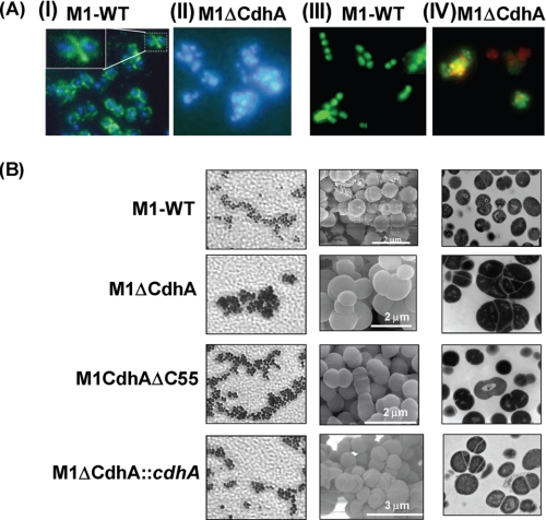
FIGURE 3.
Light, scanning, and transmission electron microscopy of M1-SF370 wild-type and M1ΔCdhA, M1CdhAΔC55, and M1ΔCdhA::cdhA mutant GAS strains. A, indirect immunofluorescence microscopy of intact wild-type SF370 and the mutant M1ΔCdhA strain using anti-CdhA antibody, FITC-labeled conjugate antibody, and DAPI (blue nuclear stain). CdhA is primarily located at the septum (green fluorescence in I) of M1-SF370. The inset in the first panel shows the magnification of a region localized with CdhA in one of the dividing bacteria. II, the M1ΔCdhA strain forming cluster and the absence of green fluorescence (only nuclear staining with DAPI). III and IV were stained with Live-Dead bacteria stain. The presence of red stain in some portion of the clusters of M1ΔCdhA (IV) indicates the presence of dead bacteria. B, images acquired for the M1-WT, M1ΔCdhA, M1CdhAΔC55, and M1ΔCdhA::cdhA strains employing light microscopy, SEM, and TEM are shown in left, middle, and right panels, respectively. The indicated scale mark applies to both SEM and TEM for each strain. Light microscopy was carried using Gram stain.

