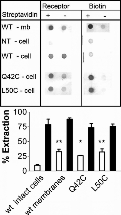
FIGURE 5.
Top, membranes from cells expressing the wild type receptor (row 1); intact, non-transfected HEK cells (row 2); or cells expressing the transiently transfected wild type (row 3), Q42C (row 4), or L50C (row 5) glucagon receptors were treated with maleimide-PEO2 biotin before streptavidin extraction. Membrane samples were solubilized and extracted in the absence (lanes 1 and 3) or presence (lanes 2 and 4) of immobilized streptavidin. The supernatants were blotted and tested for the presence of glucagon receptor (columns 1 and 2) or biotin (columns 3 and 4). Results shown are representative of 3–6 experiments. Bar graph, extraction of the glucagon receptor (gray bars) and of total biotin (used as control; white bars), expressed as a percentage of the signal observed in non-extracted samples. The average ± S.D. (error bars) of 3–6 experiments is shown. *, significantly different from WT (p < 0.05); **, significantly different from WT (p < 0.01).

