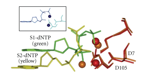
Figure 6.
Side view of a dNTP in the “chair-like” shape S1-dNTP (green) versus the “goat-tail-like” shape S2-dNTP (yellow). Key amino acids (only D7, D105, and K159 are shown) from a Dpo4 structure adopting the S1-dNTP shape were superimposed on the same amino acids in a Dpo4 structure adopting the S2-dNTP. Spheres are divalent cations (S1-dNTP/red and S2-dNTP/brown). S1-dATP from T7 DNA polymerase is also shown (insert, blue). X-ray coordinates are from 1SOM-B for Dpo4/S1-dNTP, 1RYS-A for Dpo4/S2-dNTP, and 1T7P for T7 DNAP.

