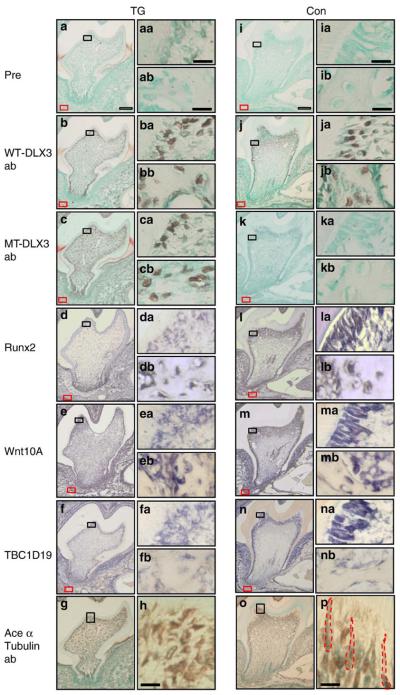
Fig. 6.
Expression patterns of WT-DLX3, MT-DLX3, and acetylated α-tubulin proteins and Runx2, Wnt10A, and TBC1D19 mRNA in the mandibular first molars of 14-day-old mice (a–h: teeth sections of TG mouse; i–p: teeth sections from control mouse). Panels a and i show prebleeding rabbit antisera; panels b and j show WT-DLX3-specificantibody; panels c and k show MT-DLX3-specific antibody; panels d and l show Runx2 in situ hybridization; panels e and m show Wnt10A in situ hybridization; panels f and n show TBC1D19 in situ hybridization; panels g, h (high-power magnification), o, and p (high-power magnification) show acetylated α-tubulin antibody. Panels aa–na show high-power magnification of differentiated odontoblasts from the black rectangular box seen in the respective low-power panels. Panels ab–nb show high-power magnification of osteoblasts of the alveolar bone from the areas indicated by a red rectangular box in the respective low-power panels. Higher-power magnification of acetylated α-tubulin staining show primary ciliary morphology of odontoblasts in control mice (red dot line in panel p) but not in TG mouse (h). White scale bars are 100 μm, and black scale bars are 10 μm.

