
Figure 1.
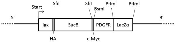
Schematic representation of the expression unit in the pDisplaySacLac2 and pBabe SacLac2 vectors. From 5′ to 3′, the unit begins at ATG start codon followed by the murine Igκ leader sequence and an HA epitope tag. Next, is the bacterial selection marker SacB is flanked by SfiI restriction sites, followed by a c-Myc epitope. This is followed by a membrane spanning segment derived from the human PDGFR gene, and then a LacZα gene flanked by PflmI restriction sites.