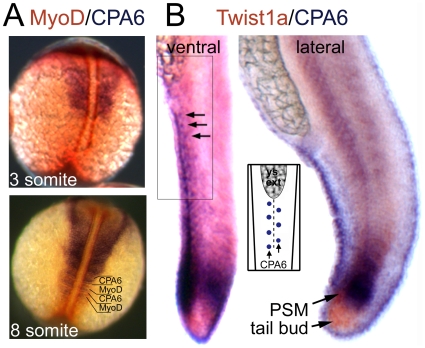
Figure 7. Distribution of CPA6 mRNA compared with somitogenesis markers.
In situ hybridization was performed with RNA probes specific for (A) CPA6 (purple) and MyoD (orange) at 3 and 8 somite stages (11 and 14 hpf), and (B) CPA6 (purple) and Twist1b (orange) at 24 hpf. Arrows indicate ectodermal cells arranged along the ventral ridge of the tail and expressing CPA6. The regular arrangement of these cells, also found along the dorsal ridge, is illustrated in the inset. PSM, presomitic mesoderm; ys ext, yolk-sac extension.

