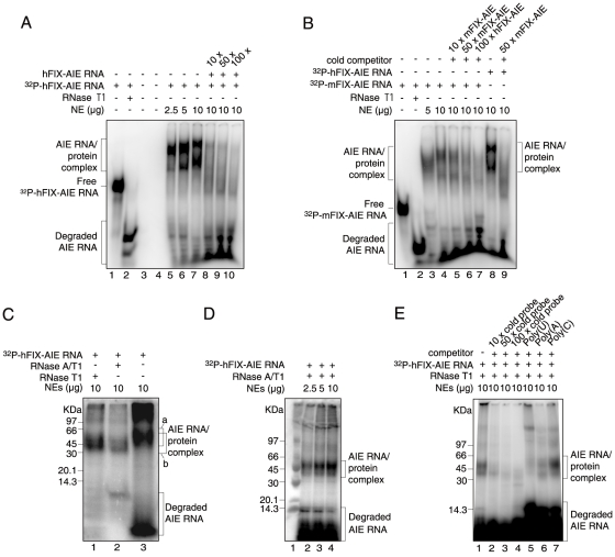
Figure 1. EMSAs and SDS-PAGE analyses of 32P-hFIX-AIE RNA/nuclear protein and 32P-mFIX-AIE RNA/nuclear protein complexes.
A. EMSAs of 32P-hFIX-AIE RNA with liver NEs. Various conditions tested are shown at the top. Brackets on the left indicate hFIX-AIE RNA probe-protein complexes and degraded 32P-hFIX-AIE RNA as shown. The position of free 32P-hFIX-AIE RNA is shown with a short horizontal bar on the left. Lanes 1, 2 and 5–10 contain 32P-hFIX-AIE RNA. Lane 2, treated with RNase T1; lanes 3 and 4, null controls; lanes 5–7, with increasing amounts of liver NEs; lanes 8–10, with NEs (10 µg) and increasing amounts of cold hFIX-AIE RNA competitor. B. EMSAs of 32P-mFIX-AIE or 32P-hFIX-AIE RNA with liver NEs. Lanes 1–7 contains 32P-mFIX-AIE RNA, while lanes 8 and 9 contain 32P-hFIX-AIE RNA. Positions of the AIE RNA probe-protein complex, free AIE RNA probe and degraded probe are similarly shown as in A. Lane 1, without NEs; lane 2, treated with RNase T1; lanes 3 and 4, with increasing amounts of NEs; lanes 5 and 6, with NEs (10 µg) and increasing amounts of cold mFIX-AIE RNA competitor; lane 7, with NEs (10 µg) and cold hFIX-AIE RNA; lane 8, with NEs (10 µg); lane 9, with NEs (10 µg) and cold mFIX-AIE RNA. C. SDS-PAGE analysis of UV cross-linked 32P-hFIX-AIE RNA/nuclear protein complex treated with RNase T1 (lane 1), RNase A/T1 (lane 2) and no RNase treatment (lane 3). Brackets a and b represent the positions of AIE RNA-protein complex without and with RNase-treatment, respectively. Size marker positions are shown on the left. D. SDS-PAGE analysis of UV cross-linked and RNase T1-treated 32P-hFIX-AIE RNA incubated with increasing amounts of NEs. Positions for the AIE RNA/protein complex and degraded 32P-hFIX-AIE RNA are shown with brackets on the right. Lane 1, protein size marker with sizes shown on the left. E. SDS-PAGE analysis after competitive EMSAs of 32P-hFIX-AIE RNA with cold hFIX-AIE RNA (lanes 2–4), and with poly(U), poly(A) and poly(C) (lanes 5–6). Other conditions are similar to D.

