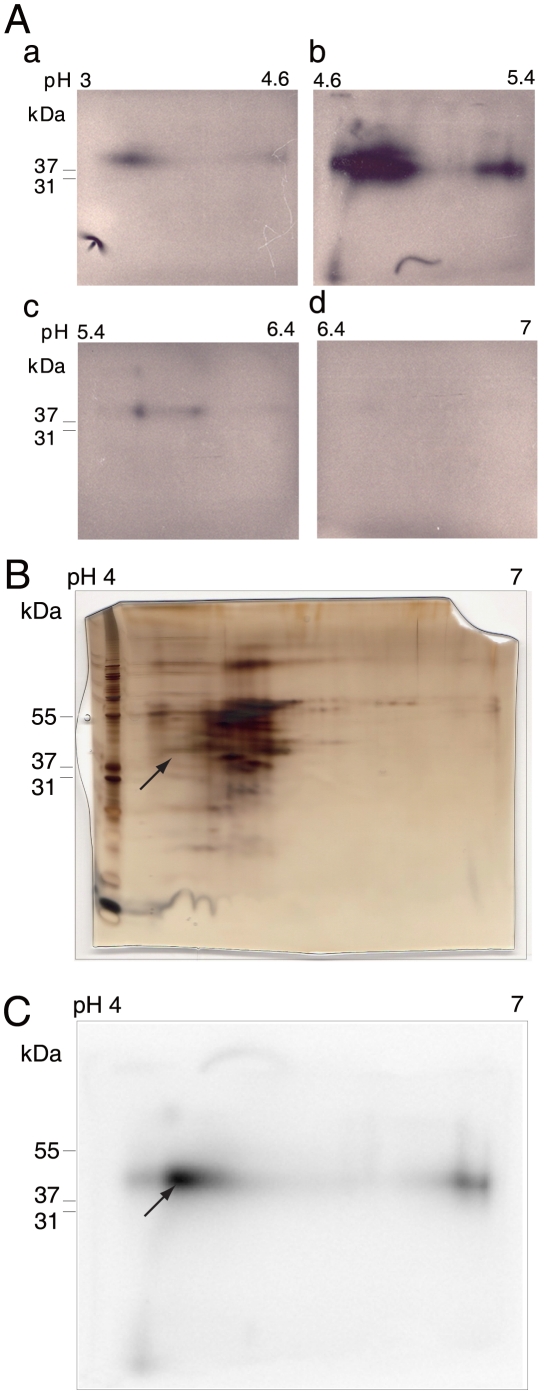
Figure 2. 2DE analysis of UV cross-linked/RNase-treated 32P-hFIX-AIE RNA/liver nuclear protein complex and its autoradiography.
A. Analytical scale 2DE analyses of the solution-phase IEF chamber solutions containing UV cross-linked/RNase-treated 32P-hFIX-AIE RNA/protein complexes. Chamber solutions of pH 3–4.6, pH 4.6–5.4, pH 5.4–6.4 and pH 6.4–7.0 zones were subjected to analytical 2DEs using immobilized pH 4–7 gradient gels for IEF and 4–12% gradient gel SDS-PAGE, and the results are shown in panels a, b, c and d, respectively. B. Silver-stained 2DE gel of UV cross-linked and RNase-treated 32P-hFIX-AIE RNA/nuclear protein complexes concentrated to pH 3–4.6 and pH 4.6–5.4 chambers in the solution phase IEF. Molecular size marker proteins are shown on the left edge of the gel with their sizes. Arrow indicates the protein spot position matching with the center of the major radioactive spot detected by autoradiography. C. Autoradiogram of the gel shown in B. Arrow indicates the center of the major radioactive spot, and the gel recovered from this center area was subjected to subsequent MALDI-TOF/MS analysis.

