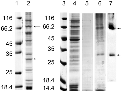
Figure 3. Purification of D-CF produced mAQP4 M23 in 0.2% Brij-35 by Co2+-NTA chromatography.
Samples were separated by 12% (lanes 1–2) or 16% (lanes 3–7) SDS-PAGE and analysed by Coomassie staining. Lanes 1 and 3, protein marker; Lane 2, precipitate after P-CF expression; Lane 4, flow through; Lane 5, washing fraction; Lane 6, elution fraction; Lane 7, immunoblot of lane 6 using anti-His antibodies. Samples of 2 µl were applied to each lane.

