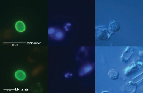
Fig. 2.
Micrographs of typical Cryptosporidium oocysts confirmed by fluorescence microscopy using differential interference contrast optics, found in 2 intake water samples (up and down) from the Han River. (Left) An oocyst showing its brilliant apple-green fluorescence with ovoid shape, stained with mAb, 5 µm in diameter under a blue filter; (Middle) 1-4 blue points by DAPI nuclei staining under a UV filter; (Right) 1-4 sporozoites shown by DIC.

