Figure 3.
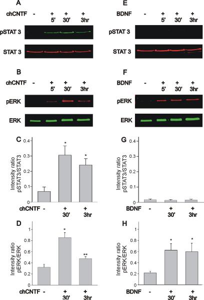
Time course of STAT3 and ERK activation in nodose sensory neurons following stimulation with chCNTF or BDNF. A, B) Stimulation of nodose cell cultures with chCNTF generates a considerable increase in STAT3 and ERK phosphorylation. C) Phosphorylation pattern of STAT3 as determined by the intensity ratio of pSTAT3 to total STAT3. Stimulation of nodose neurons with chCNTF for 30 min causes a significant increase in the pSTAT3/STAT3 ratio. D) Phosphorylation pattern of ERK as determined by the intensity ratio of pERK to total ERK. Note that stimulation with chCNTF for 30 min causes a significant increase in the pERK/ERK ratio. The pERK/ERK intensity ratio decreases significantly after 3 hr stimulation with CNTF (n=8). E, F) Stimulation of nodose cell cultures with BDNF results in a significant increase in ERK activation without evoking STAT3 activation. G) The intensity ratio of pSTAT3 to total STAT3 was very low following stimulation of nodose neurons with BDNF. Notice that the scale of the Y-axis in figures C and G are the same in order to visualize differences in the pattern of STAT3 activation evoked by chCNTF and BDNF. H) As determined by the pERK/total ERK intensity ratio, BDNF stimulation of nodose neurons causes a significant increase in ERK phosphorylation, that was maintained for up to 3 hr (n=7). Cell cultures of nodose neurons were treated with chCNTF (50 ng/mL) or BDNF (50 ng/mL) for various lengths of time (5 min, 30 min and 3 hr). Cell lysates were collected and subjected to immunoblot analysis using a two-color western blot detection with the Odyssey infrared imaging system. * denotes p ≤ 0.05 vs. control (no treatment), ** denotes p ≤ 0.05 vs. CNTF treatment for 30 min.

