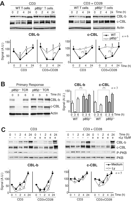
Figure 4.
Defective CBL and TCR down-regulation in p85β-deficient cells. (A) Purified peripheral T cells from p85β+/− and p85β−/− mice were stimulated with CD3+CD28 Ab for the indicated times. CBL-b and c-CBL levels were analyzed in WB. Actin was used as loading control. (B) For primary responses, p85β+/− and p85β−/− F5TCRTg mice were injected with antigenic peptide. Mice were sacrificed at 0, 3, and 6 hours after injection and CBL levels were examined in extracts of purified T cells. (C) Jurkat T cells were activated with CD3 or CD3 + CD28 for the indicated times in the presence or absence of 10 μM Ly294002. We analyzed CBL-b and c-CBL levels in cell extracts by WB. Graphs (A-C) show CBL-b and c-CBL signal intensity in AUs, mean plus or minus SD (n = 5). *P < .05.

