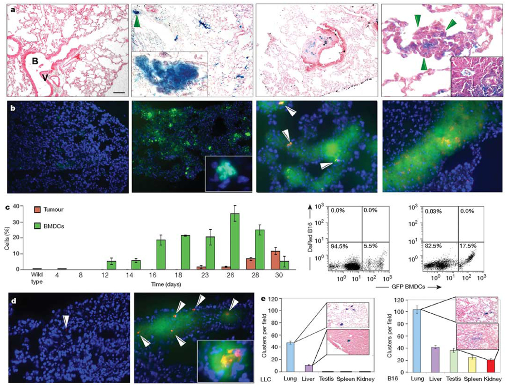
Figure 1. Bone marrow-derived cells form the pre-metastatic niche.
a, β-gal+ bone marrow cells (left panel) are rarely observed in lungs after irradiation and before LLC cell implantation (n = 6). By day 14, β-gal+ bone marrow-derived clusters appear in the lung parenchyma (left middle panel and magnified inset of the region arrowed; n = 25) and are associated with micrometastases by day 23 (right panel, arrows) and in gross metastases (right panel, inset; n = 12). Also shown is a cluster with associated stroma between a terminal bronchiole and bronchial vein, a common metastatic site (right middle panel). B, terminal bronchiole; V, bronchial vein. b, GFP+ bone marrow in the lungs after irradiation and before DsRed-tagged B16 cell implantation (left panel; n = 6). On day 14, GFP+ (green) BMDCs are seen with no DsRed+ (red) tumour cells (left middle panel and inset; n = 12). Beginning on day 18, a few single DsRed+ B16 cells adhere to GFP+ bone marrow clusters (right middle panel), and by day 23, DsRed+ tumour cells proliferate at cluster sites (right panel; n = 8). DAPI stain (blue) shows cell nuclei. c, A graph showing flow cytometric data of bone marrow-derived GFP+ BMDCs and DsRed+ B16 cells in the lung, and two flow diagrams on day 14 (left panel) and day 18 (right panel) (n = 30; error bars show s.e.m.). d, GFP+ BMDCs mobilized with B16 conditioned media, then DsRed-tagged tumour cells injected through the tail vein adhere 24 h later (right panel, arrows) compared with animals receiving media alone (left panel; P < 0.01). Inset shows proliferating tumour cells in a cluster after four days (right panel inset; n = 6). e, Number of clusters per ×100 objective field in animals with intradermal LLC or B16 tumours (n = 12). Scale bar on top left panel applies to panels a (left, left middle, right middle, 80 µm; left middle inset, 8 µm; right, 20 µm; right inset, 47 µm), b (left, left middle, 80 µm; left middle inset, 8 µm; right middle, right, 40 µm) and d (40 µm; right inset, 20 µm).

