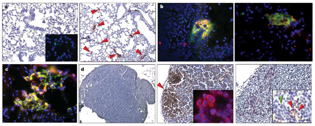
Figure 2. Pre-metastatic clusters are comprised of VEGFR1+ haematopoietic progenitors.
a, VEGFR1 staining in irradiated lung before tumour implantation (left panel and inset; n = 10) and 14 days after LLC cell implantation showing clusters in the lung (right panel, arrows; n = 18, 3.9 ± 0.2% cells with VEGFR1 staining per ×100 objective field, P < 0.05). b, c, Double immunofluorescence in the lung of an animal with day 14 LLC tumour. b, VEGFR1+ (red) and GFP+ (green) bone marrow cells (left panel), VEGFR1+ (red) and CD133+ (green) (right panel). c, VEGFR1+ (red) and CD117+ (green). d, VEGFR1+ clusters in c-Myc transgenic lymph node at day 40 of life and before tumorigenesis (middle panel and inset showing VEGFR1+ cells (red)) as compared with wild-type littermate lymph node without the transgene (left panel), and day 120 c-Myc transgenic node with lymphoma (right panel). In the inset of the right panel, arrows indicate the VEGFR1+ clusters (red) surrounded by lymphoma (green) (n = 6). Scale bar at bottom right applies to panels a (80 µm; left inset, 40 µm), b (20 µm), c (20 µm) and d (80 µm; insets, 8 µm).

