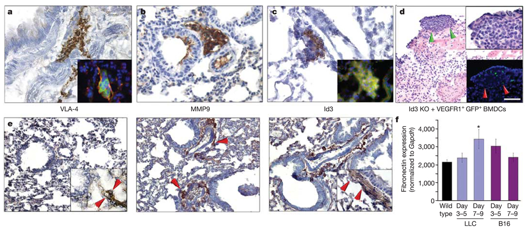
Figure 5. The VLA-4/fibronectin pathway mediates cluster formation.
a–c, Wild-type mice 14 days after tumour implantation develop clusters expressing VLA-4 (inset, VEGFR1 (red) and VLA-4 (green)), MMP9 and Id3 (inset, VEGFR1 (red) and Id3 (green)). d, Lung tissue in Id3 knockout (KO) mice with LLC tumours given VEGFR1+GFP+ BMDCs (P < 0.01 by ANOVA; n = 6). Green arrows show region in upper inset. Red arrows (lower inset) show the site of metastasis with GFP+VEGFR1+ cells. e, Baseline fibronectin expression in the wild-type lung (n = 6) (left panel). Increased stromal fibronectin in the peribronchial region of the pre-metastatic lung at day three (middle panel, arrows), with maximal expression on day 14 (right panel). Insets, PDGRFα expression indicates resident fibroblasts laying down fibronectin. f, Quantitative RT–PCR reveals increased fibronectin expression in the lungs of mice with LLC tumours compared with wild type (*P < 0.05 by ANOVA; n = 6), and a similar earlier trend in lungs from animals with B16 melanoma. Scale bar at top right applies to panels a, b, c (40 µm; insets, 8 µm), d (80 µm; top right inset, 20 µm; bottom right inset 80 µm) and e (40 µm; insets, 20 µm).

