Figure 2.
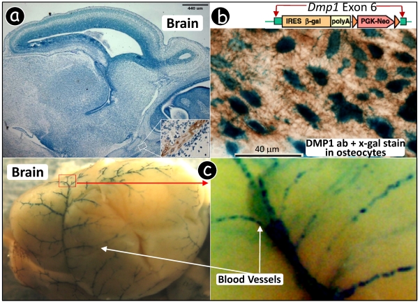
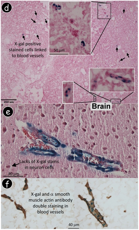
Dmp1 lacZ knock-in transgene is expressed in blood vessels. (a) Immunohistochemistry using a polyclonal antibody against DMP1 N-terminal showed DMP signal in bone only. (b) Dmp1 exon 6, containing 80% of most coding region, is replaced by a lacZ reporter gene whose expression reflects endogenous DMP1 expression pattern. A decalcified frozen section of alveolar bone was stained with X-gal followed by DMP1 antibody immunostaining, showing a blue stain in nuclei of osteocytes (lacZ expression) and brown stain in matrix (DMP1 expression). (c) Whole mount X-gal staining of a heterozygous Dmp1-lacZ knock-in brain overnight showed blue stain in blood vessels (3-wk old, left panel), and the signal appears in a particular group of muscle cells (right panel, the enlarged area in the left panel). (d) A frozen section of brain tissue was stained with X-gal, showing the blue stained cells which are similar to pericytes. (e) Whole mount X-gal stain of brain overnight was followed by paraffin section with an H&E count stain, showing blue stained cells in blood vessels but not in the neural cells. (f) A double stained image with an X-gal stain followed by an anti-muscle actin antibody stain showed that all muscle cells in the blood vessels were immunoreactive to the antibody but the blue-stained cells were limited in a certain type of cells.


