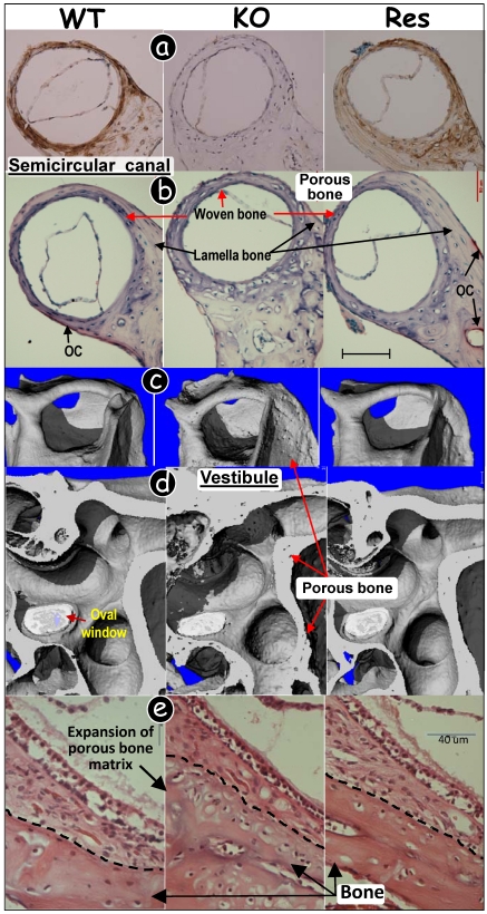
Fig 4.
Targeted expression of DMP1 rescued malformed inner ear bone in Dmp1-null mice. (a) Immunohistochemistry assay showed that DMP1 was highly expressed in 3-wk-old WT (left panel, brown color) and rescued (Res, targeted-reexpression of DMP1 in Dmp1 KO background, right panel) semicircular canal bones with no signal in KO bone (middle panel). (b) TRAP stained images showed few osteoclasts (OC) and a reduction of lamella bone volume in the KO semicircular canal (middle panel), and these pathological changes were restored in the Res canal (right panel) compared to the WT group (left panel). (c-d) μCT images showed porous bone in the KO semicircular canal (c), and vestibular organ (d)(middle panels); and this change was restored in all Res bones (right panels) compared to the WT control (left panels). (e) H&E stained images showed expanded bony mass in the KO vestibular apparatus, and this change was fully restored in the Res bone (right panel) compared to the WT control (left panel).

