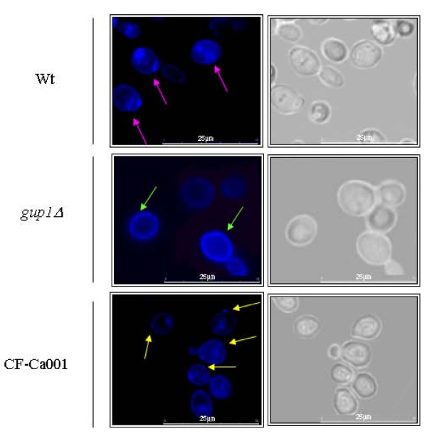
Figure 2.
Sterol lipid distribution is affected by the deletion of CaGUP1 mutation. The images show filipin staining of the wt, Cagup1Δ null mutant and CF-Ca001 strain cells grown in YPD till mid-exponential phase. Cells were stained with a fresh solution of filipin (5 mg/ml), stabilized onto slides with a drop of an anti-fading agent, and promptly visualized and photographed. Pink and yellow arrows point to punctuated filipin stained sterols at the level of plasma membrane in the wt and CF-Ca001 strains respectively. Green arrows point to filipin stained sterols evenly distributed in the Cagup1Δ null mutant plasma membrane. The gup1Δ photos are representative of the results obtained with the several clones (3-5) of Cagup1Δ null mutant strain tested.

