Figure 6.
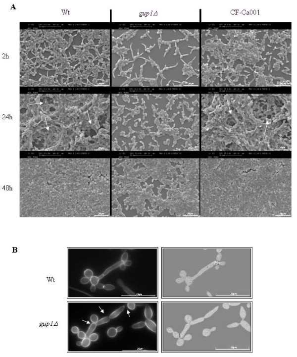
Cagup1Δ null mutation causes less and differently structured time-course biofilm formation. (A) SEM micrographs of time course biofilm formation. Arrows indicate the channels observed in a typical biofilm structure - wt and CF-Ca001- not observed in Cagup1Δ null mutant strain biofilm. (B) Chitin assembly by CFW staining of individual cells observed by LM. Distinct filament types can be observed. Wt cells display hyphae without septae constrictions, the first septum located within the germ tube, apart from the mother-bud neck (arrow), and less branched, thinner elongated compartments with parallel sides. Cagup1Δ null mutant strain cells present pseudohyphae with constrictions located at the septae junctions and at the mother-bud neck, where the first septum is located (arrows), highly branched and thicker elongated compartments without parallel sides. The gup1Δ photos are representative of the results obtained with the several clones (3-5) of Cagup1Δ null mutant strain tested.

