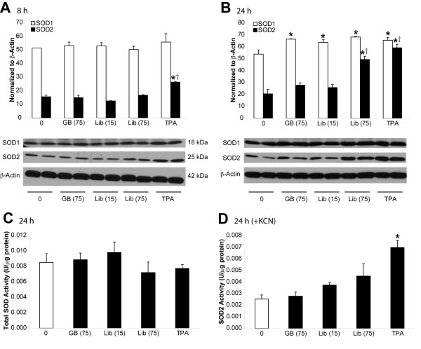
Figure 2.
Effects of Libby six-mix exposure on SOD levels as demonstrated by Western blots and activity assays. Immunoblotting of SOD1 and SOD2 protein levels using LP9/TERT-1 cell lysates collected following (A) 8 h or (B) 24 h exposures to glass beads (non-pathogenic control), Libby six-mix, or 100 ng/ml TPA (positive control). Surface area (x106 μm2/cm2) is represented in parentheses following mineral name. A total of 30 μg of protein was separated by 10% SDS-PAGE, transferred to nitrocellulose, and immunoblotted using specific SOD1, SOD2, and β-Actin primary antibodies. Quantitative densitometry was performed using QuantityOne software, and values were normalized to β-Actin protein levels. Bars denote the mean ± SEM of n = 2 samples per group and data are representative of 3 separate experiments. * p < 0.05 compared to medium control (0). † p < 0.05 compared to glass beads. (C) Total SOD and (D) SOD2 activity in LP9/TERT-1 cells following exposure to glass beads, Libby six-mix, or 100 ng/mL TPA for 24 h as determined using the Cayman Superoxide Dismutase Assay Kit. Addition of 3 mM KCN (inhibitor of SOD1 and SOD3) allowed SOD2 activity to be assayed specifically. Total protein concentrations were determined using the Bio-Rad Protein Assay so that final SOD activities could be represented as Units/μg protein. Bars denote the mean ± SEM of n = 3 samples per group, and data are representative of 3 separate experiments. * p < 0.05 compared to all other groups.

