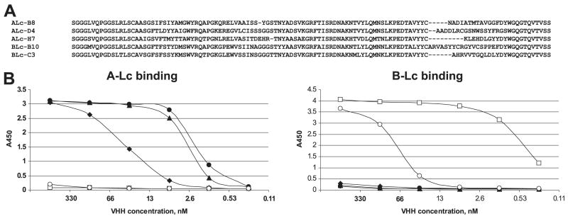
Fig. 1.
Characterization of the five selected VHH anti-BoNT Lc binding proteins. (A) Amino acid sequences of the five selected anti-BoNT Lc VHH domains aligned for maximum homology. (B) Dilution ELISA to measure binding of five VHHs to plates coated by 5 ug/ml of either BoNT/A-Lc (left) or B-Lc (right). VHH-B8 (▲), VHH-H7 (●), VHH-D4 (■), VHH-B10 (□), VHH-C3 (○). The x axis represents the VHH concentration and the y axis represents the ELISA signal.

