
Figure 2.
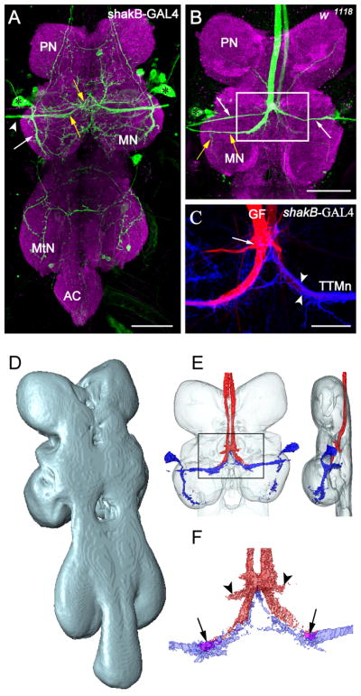
Registration of GFS components into the ventral nerve cord neuropil standard. (A) Projection view from a confocal image stack of the ventral nerve cord (VNC) showing the GFP expression pattern under the control of shakB-GAL4 (green). The neuropil was counterstained with anti-Brp (magenta). The positions of TTMn (Tergotrochanteral Motorneuron) somata are marked with asterisks. Fine processes and small somata were visible in the pro- and metathoracic neuromere (PN, MtN) and the abdominal center (AC). The TTMn extended its medial dendrite (yellow arrows) to the midline of the VNC. The posterior dendrite (white arrow) projected more ventrally in the MN. The TTMn axon (arrowhead) left the MN via the posterior dorsal mesothoracic nerve. Scale bar=50μm. (B) Projection view through the PN and MN of a wild type animal. The left Giant Fiber was dye injected with Neurobiotin (green). Dendrites and axons of the Peripherally Synapsing Interneurons (PSI, white arrows) and the TTMn (yellow arrows), and the TTMn soma (asterisk) were dye coupled to the GF. The left GF was stained due to electrical coupling of both GFs via the giant commissural interneurons in the brain. Scale bar=50μm. (C) Double staining of the GF-TTMn synaptic contact area. GFP expression under the control of shakB-GAL4 labeled the postsynaptic TTMn terminal (blue). The left GF was stained with Neurobiotin in the same preparation (red). The TTMn dendrite (arrow heads) enwrapped the GF terminal partially. The arrow marks the contact area to the PSIs. Scale bar=20μm. (D) Dorsolateral view on the surface reconstruction of the ventral nerve cord neuropil standard. (E) The projections of six reconstructed GF staining and six TTMn labels were transformed into the VNC standard and subsequently averaged. The averaged GF (red) and the averaged TTMn (blue) were displayed together as a surface reconstruction within the neuropil standard (transparent) from dorsal (left) and lateral (right). (F) Close up from the averaged GF terminal. The averaged pre- and postsynaptic terminals were aligned to each other as seen for double staining (see C). The terminals overlapped in a small area in the neuropil standard (arrows). The connection from the GF to the PSI was visible after averaging (arrowheads).