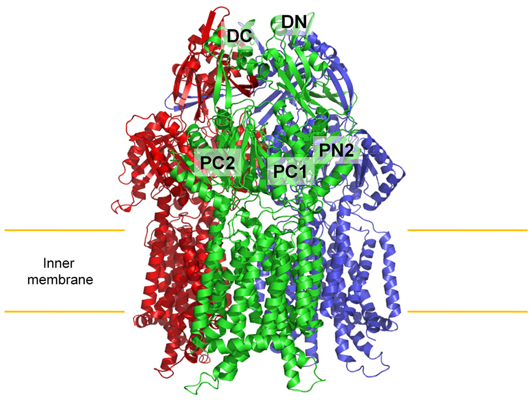
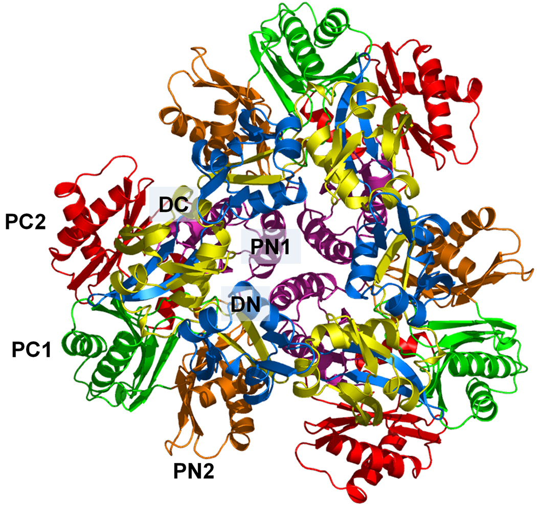
Figure 1.
Structure of the apo CusA efflux pump. (a) Ribbon diagram of the CusA homotrimer viewed in the membrane plane. Each subunit of CusA is labeled with a different color. Sub-domains DN, DC, PN2, PC1 and PC2 are labeled on the front protomer (green). The location of PN1 in this protomer is behind PN2, PC1 and PC2 (see text). (b) Top view of the CusA trimer. The six sub-domains are labeled blue (DN), yellow (DC), pink (PN1), orange (PN2), green (PC1) and red (PC2). In the apo-CusA structure, the cleft between PC1 and PC2 is closed.


