Figure 4.
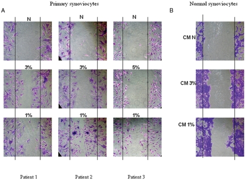
Migration assays in primary and normal synoviocytes. A. Representative images of primary synoviocytes wound repair assays exposed to normoxia (n=3), in vivo oxygen levels (n=2 at 3%, n=1 at 5%), and positive control of 1% oxygen for 24 h. Significant migration was observed when primary synoviocytes (patients 1 and 2) were exposed to 3% when compared to normoxia (A, left and middle panels), an effect that was greater at 1% hypoxia. When primary synovial fibroblast cells (SFCs) were exposed to 5% hypoxia (patient 3) migration was minimal and similar to normoxia (A right panel), migration was demonstrated when exposed to 1%. B. Normal synoviocytes cultured in conditioned supernatant from primary SFCs exposed to normoxia, 3% and 1% oxygen for 24 h before scratches. Significant migration was observed in those incubated with conditioned media from 1% and 3% hypoxic conditions. CM, cultured media; N, normoxia.

